 (ACS).
According to the ACS, cancer is a group of diseases characterized by
uncontrolled growth and spread of abnormal cells.
If the spread is not controlled, it can result in death.
(ACS).
According to the ACS, cancer is a group of diseases characterized by
uncontrolled growth and spread of abnormal cells.
If the spread is not controlled, it can result in death.Cancer WebQuest
The Cancer WebQuest introduces you to the history of diagnosing and treating cancer, basic facts about the disease, how it is diagnosed and how we have been fighting cancer in our times.
In this webquest you will learn how to:
Since the earliest medical records were kept, cancer as a disease has been described in the history of medicine. The earliest known descriptions of cancer appear in seven papyri, discovered and deciphered late in the 19th century. They provided the first direct knowledge of Egyptian medical practice. Two of them, known as the "Edwin Smith" and "George Ebers" papyri, contain descriptions of cancer written around 1600 B.C., and are believed to date from sources as early as 2500 B.C. The Smith papyrus describes surgery, while the Ebers' papyrus outlines pharmacological, mechanical, and magical treatments.
Based on the information recorded on papyri and hieroglyphic inscriptions, ancient Egyptians were able to distinguish benign tumors from malignant tumors. They were also able to use different treatments, including surgery, and other various modes of medicine.
Following the decline of Egypt, the next chapters of medical and scientific history were written in Greece and Rome. The great doctors Hippocrates and Galen dominated medical thought for 1500 years. They lifted medicine out of the realms of magic, superstition, and religion. Hippocrates and Galen defined disease as a natural process, and based treatment on observation and experience. Cancers were identified, with warnings against treatment of the more severe forms. Hippocrates is credited with naming "cancer" as "karkinoma" (carcinoma) because a tumor looked like a "crab" ("karkinoma" is Greek for "crab") in that there is a central body to a tumor and the tumor extension appeared as the legs of the "crab".
After the fall of Rome, Constantinople became the intellectual storehouse of civilization. From there, in Arabic translations, classic Greek and Roman texts made their way back through Europe. The ancient teachings of Galen continued to inspire physicians in Constantinople, Cairo, Alexandria, Athens, and Antioch in a time when magic spells and myths dominated the West. Cancer continued to be explained as the result of an excess of black bile, curable only in its earliest stages.
In the modern world, science and surgery advanced as physicians returned to direct observation of the human body. However, the theory that cancer was caused by an excess of black bile continued to prevail in the 16th century. Cancer was considered incurable, although a wide variety of pastes containing arsenic were formulated to treat its manifestations. In the 17th century, the old theory of disease based on bodily humors was discarded when Gaspare Aselli discovered the vessels of the lymphatic system and suggested abnormalities of lymph as the primary cause of cancer.
Rejecting the 17th-century theory about the cause of cancer was the French physician Claude Gendron. He concluded that cancer arises locally as a hard, growing mass, untreatable with drugs, and must be removed with all its "filaments."
Two 18th-century French scientists, physician Jean Astruc and chemist Bernard Peyrilhe, conducted experiments to confirm or disprove hypotheses related to cancer. Their efforts, however absurd they seem in retrospect, established experimental oncology, the science of seeking better diagnosis, treatments and understanding of the causes of cancer. During this period, environmental cancers were reported, and hospitals specializing in cancer care were opened.
In the late 19th century, the development of better microscopes not only helped document and define disease-causing organisms, but also made possible the examination of cells and cellular activity. Study of cancer tissues and tumors revealed that cancercells were markedly different in appearance than normal cells of surrounding tissue or the cells from which they originated. Researchers began to focus on questions such as the origin of cells and the relationship of disease to the behavior of a cell. It was the invention of the microscope that revealed the cancer cell itself.
The early 20th century saw great strides made in understanding the structures, functions and chemistry of living organisms. Cancer research in cell culture, chemical carcinogens, diagnostic techniques and chemotherapy firmly established oncology as science. Researchers pursued different theories of the origin of cancer, subjecting their hypotheses to systematic experimentation. A viral cause of cancer in chickens was documented in 1911, and both chemical and physical carcinogens were conclusively identified. Chromosomal abnormalities were also investigated as possible causes of cancer.
In 1913, a need to combat rising public fear and ignorance concerning cancer led to two significant events: the publication of the first known article on cancer's warning signs in a popular woman's magazine, and formation of a nationwide organization dedicated to public education on cancer. Cancer, as a disease, was brought into the light of day.
In 1937, the U.S. Congress made the conquest of cancer a national goal with a unanimous vote to pass the National Cancer Institute Act. This Act created the National Cancer Institute, which was expected to break new theoretical ground by conducting its own research, promoting research in other institutions and coordinating cancer-related projects and activities. In 1971, President Richard M. Nixon signed the National Cancer Act, launching a National Cancer Program administered by the National Cancer Institute. Key events in the United States' national cancer policy legislative history, from 1937 to 1999 are available here.
Since its establishment, fundamental biomedical research supported by the National Cancer Institute has advanced the understanding of cancer. Using tools of molecular biology and molecular genetics, scientists are making great leaps in the discovery and mapping of links between chromosomes, the genes within, and cancer. In addition to traditional cancer therapies, potential solutions to the prevention and cure of cancer seem limited only by the imagination.
There are many texts and references
that attempt to define cancer.
The simplest definition is from the American
Cancer Society (ACS).
According to the ACS, cancer is a group of diseases characterized by
uncontrolled growth and spread of abnormal cells.
If the spread is not controlled, it can result in death.
Cells are the structural units of all living things. Each of us has trillions of cells, as does a growing tree. Cells make it possible for us to carry out all kinds of functions of life: the beating of the heart, breathing, digesting food, thinking, walking, and so on. However, all of these functions can only be carried out by normal healthy cells. Some cells stop functioning or behaving as they should, serving no useful purpose in the body at all, and become cancerous cells.
The most fundamental characteristic of cells is their ability to reproduce themselves. They do this simply by dividing: one cell becomes two, the two become four, and so on. The division of normal and healthy cells occurs in a regulated and systematic fashion. In most parts of the body, the cells continually divide and form new cells to supply the material for growth or to replace worn-out or injured cells. For example, when you cut your finger, certain cells divide rapidly until the tissue is healed and the skin is repaired. They will then go back to their normal rate of division. In contrast, cancer cells divide in a haphazard manner. The result is that they typically pile up into a non-structured mass or tumor.

Sometimes tumors do not stay harmlessly in one place. They destroy the part of the body in which they originate and then spread to other parts where they start new growth and cause more destruction. This characteristic distinguishes cancer from benign growths, which remain in the part of the body in which they start. Although benign tumors may grow quite large and press on neighboring structures, they do not spread to other parts of the body. Frequently, they are completely enclosed in a protective capsule of tissue and they typically do not pose danger to human life like malignant tumors (cancer) do.
Although cancer is often referred to as a single condition, it actually consists of more than 100 different diseases. These diseases are characterized by uncontrolled growth and spread of abnormal cells. Cancer can arise in many sites and behave differently depending on its organ of origin. Breast cancer, for example, has different characteristics than those of lung cancer. It is important to understand that cancer originating in one body organ takes its characteristics with it even if it spreads to another part of the body. For example, metastatic breast cancer in the lungs continues to behave like breast cancer when viewed under a microscope, and it continues to look like a cancer that originated in the breast.
The word cancer comes from the Latin (originally Greek) derived term for crab, because of the way a cancer adheres to any part that it seizes upon in an obstinate manner like the crab. Hippocrates first described cancer as having a central body with the tendency to reach out and spread like "the arms of a crab." Besides the popular, generic term "cancer" used by most people, there is another more technical term: neoplasia. Neoplasia (neo = new, plasia = tissue or cells) or neoplasm literally means new tissue in Greek. This indicates that cancers are actually new growths of cells in the body.
Another term for cancer is "malignant tumor." Tumor literally means "swelling" or "mass." In this case, it refers to a mass of non-structured new cells, which have no known purpose in the physiological function of the body.
There are two general types of tumors: benign (non-cancerous) tumors and malignant (cancerous) tumors. A benign tumor is composed of cells that will not invade other unrelated tissues or organs of the body, although it may continue to grow in size abnormally. A malignant tumor is composed of cells that invade the basement membrane and invade or spread to other parts of the body. This occurs either by direct extension to neighboring organs and/or tissues or by metastasizing to distant sites by means of the vascular system (the blood stream), the lymphatic system, or by seeding or implantation of cancer cells in body cavities.
Terms such as "mass" and "lump" are used to describe any overgrowth of tissue. However, these terms may not necessarily mean that such growths contain cancer cells.
In addition to neoplasia, there are several other terms referring to abnormal cell growth. These include the following:
Hyperplasia refers to an abnormal increase in the number of cells, which are in a normal component of that tissue and are arranged in a normal fashion with subsequent enlargement of the affected part. One example is thyroid hyperplasia, an enlargement of the thyroid gland caused by an abnormal rapid growth of the epithelial cells lining the follicles. Another example is: Guitar strumming leads to hyperplasia of the cells on the thumb (a callus is formed). The callus on the thumb is a hyperplastic growth.
Hypertrophy refers to an abnormal increase in the size of each cell, for example, the increase in cell size of cardiac muscle.
Metaplasia refers to the replacement of one mature cell type with another mature cell type: for example, squamous metaplasia of the respiratory columnar epithelium — as evidenced by the metaplastic cough of a smoker.
Dysplasia refers to the replacement of one mature cell type with a less mature cell type: for example, dysplasia of the cervix epithelium.
Hyperplasia, metaplasia, and dysplasia are reversible because they are results of a stimulus. Neoplasia is irreversible because it is autonomous.
Names of benign tumors usually end with "oma" regardless of their cell type. For example, a benign glandular tumor (epithelium tissue) is called adenoma and a benign bone tumor is called osteoma, while a malignant glandular tumor is called adenocarcinoma and a malignant bone tumor is called osteosarcoma.
In addition to benign tumors, there are in situ tumors and invasive tumors. In situ tumors do not invade the basement membrane, whereas invasive tumors do invade the basement membrane.

The cell is the fundamental unit of life. It is the smallest structure of the body capable of performing all of the processes that define life. Each of the organs in the body, such as the lung, breast, colon, and brain, consists of specialized cells that carry out the organ's functions such as the transportation of oxygen, digestion of nutrients, excretion of waste materials, locomotion, reproduction, thinking, etc.
To assure the proper performance of each organ, worn out or injured cells must be replaced, and particular types of cells must increase in response to environmental changes. For example, the bone marrow increases its production of oxygen-carrying red blood cells sevenfold or greater in response to bleeding or high altitude. Certain white blood cells are produced more rapidly during an infection. Similarly, the liver or endocrine organs frequently respond to injury by regenerating damaged cells.
As stated in the previous section, reproduction of cells is a process of cell division. The division of normal cells is a highly regulated process. The cell growth, inheritance and containment is controlled by its DNA (deoxyribonucleic acid).
DNA is a highly complex molecule manufactured in the cell nucleus and serves as the cell's "brain." DNA is the blueprint for everything the cell does. In a human cell, the DNA is arranged in 46 distinct sections called chromosomes. They are arranged in pairs, 23 chromosomes from each biological parent.
Together, the 46 chromosomes contain more than 100,000 genes. A gene is a segment of DNA that determines the structure of a protein, which is needed for development and growth as well as carrying out vital chemical functions in the body. Like the chromosomes, genes are arranged in pairs — one gene from the mother and one from the father.
Each gene occupies a specific location on a chromosome. Through a number of biochemical steps, each gene tells a cell to make a different protein. Some genes instruct the cell to manufacture structural proteins, which serve as building blocks. Other genes tell the cell to produce hormones, growth factors or cytokines, which exit the cell and communicate with other cells. Still other genes tell the cell to produce regulatory proteins that control the function of other proteins or tell other genes when to turn "on" or "off." When a gene is turned on, it manufactures another complex molecule called ribonucleic acid (RNA), which contains all the information the cell needs to make new proteins.
Cells divide only when they receive the proper signals from growth factors that circulate in the bloodstream or from a cell they directly contact. For example, if a person loses blood, a growth factor called erythropoietin, which is produced in the kidneys, circulates in the bloodstream and tells the bone marrow to manufacture more blood cells.
When a cell receives the message to divide, it goes through the cell cycle, which includes several phases for the division to be completed. Checkpoints along each step of the process make sure that everything goes the way it should.
Many processes are involved in cell reproduction and all these processes have to take place correctly for a cell to divide properly. If anything goes wrong during this complicated process, a cell may become cancerous.
A cancer cell is a cell that grows out of control. Unlike normal cells, cancer cells ignore signals to stop dividing, to specialize, or to die and be shed. Growing in an uncontrollable manner and unable to recognize its own natural boundary, the cancer cells may spread to areas of the body where they do not belong.
In a cancer cell, several genes change (mutate) and the cell becomes defective. There are two general types of gene mutations. One type, dominant mutation, is caused by an abnormality in one gene in a pair. An example is a mutated gene that produces a defective protein that causes the growth-factor receptor on a cell's surface to be constantly "on" when, in fact, no growth factor is present. The result is that the cell receives a constant message to divide. This dominant "gain of function gene" is often called an oncogene (onco = cancer).
The second general type of mutation, recessive mutation, is characterized by both genes in the pair being damaged. For example, a normal gene called p53 produces a protein that turns "off" the cell cycle and thus helps to control cell growth. The primary function of the p53 gene is to repair or destroy defective cells, thereby controlling potential cancerous cells. This type of gene is called an anti-oncogene or tumor suppressor gene. If only one p53 gene in the pair is mutated, the other gene will still be able to control the cell cycle. However, if both genes are mutated, the "off" switch is lost, and the cell division is no longer under control.
Abnormal cell division can occur either when active oncogenes are expressed or when tumor suppressor genes are lost. In fact, for a cell to become malignant, numerous mutations are necessary. In some cases, both types of mutations — dominant and recessive — may occur.
A gene mutation may allow an already abnormal cell to invade the normal tissue where the cancer started or to travel in the bloodstream (metastasize) to remote parts of the body, where it continues to divide.
A normal cell can become damaged in different ways. A cell can become abnormal when part of a gene is lost (deleted), when part of a chromosome is rearranged and ends up in the wrong place (translocation), or when an extremely small defect occurs in the DNA, which results in an abnormal DNA "blueprint" and production of a defective protein occurs.
Abnormal cell division can also be caused by viruses. In this case, genes may be normal, but the protein may not function normally because the cell contains a cancer-producing virus.
How a specific cancer cell behaves depends on which processes are not functioning properly. Some cancer cells simply divide and produce more cancer cells, and the tumor mass stays where it began. Other cancer cells are able to invade normal tissue, enter the bloodstream, and metastasize to a remote site in the body.
In summary, cancer cells have defects in normal cellular functions that allow them to divide, invade the surrounding tissue, and spread by way of vascular and/or lymphatic systems. These defects are the result of gene mutations sometimes caused by infectious viruses.
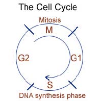
Cell division is the process by which cells reproduce (mitosis). The cell cycle is a series of changes the cell goes through from the time it is first formed until it divides into two daughter cells. It starts at mitosis (M-phase) and ends with mitosis. In between are the G-1, S, and G-2 phases. The duration of S, M and G-2 are relatively constant in different tissues.
Between the M-phase and the S-phase is a gap (G-l) where production of RNA, proteins, and enzymes needed for DNA synthesis occurs. The duration of G-1 varies and determines the length of the cell cycle.
The S-phase is when DNA synthesis occurs.
Between the S-phase and M-phase is a second gap (G-2). Cells are thought to prepare for mitosis in G-2 when specialized proteins and RNA are produced. G-0 is a dormant phase.
The four phases of mitosis are:
The search for cause(s) of cancer has been going on for centuries. Early researchers said that cancer was a natural result of aging. As cells degenerated, it was believed that some simply became malignant. Others said cancer was hereditary, and investigations into genetics began. Then some began to consider chemical links while still others questioned whether viruses or bacteria were at fault. Finally, the "irritation" theory became popular,and researchers began trying to identify irritants — such as tobacco and coal tar — that would cause cancer in laboratory animals. Ultimately, though, cancer experts were forced to confront the fact that although all these factors might be involved, none of them invariably cause cancer. Not every animal or person exposed to an irritant or a particular chemical in the laboratory developed cancer, nor did all elderly people or everyone with a family history of cancer get it. As a result, scientists had to abandon the theory that cancer had a single cause.
However, despite the fact that there is yet no absolute agreement among the cancer research community in terms of what actually causes cancer, scientists are certain that many factors can be linked to cancer. These factors, including many other possible causes of cancer suggested by cancer researchers, are believed to be "cancer risk factors." These risk factors include eating habits, lifestyle, living or working environments, genetics, and many others. Following are some major cancer risk factors identified by researchers with the support of scientific statistics:
Cigarette smoking alone is directly related to at least one-third of all cancer deaths annually in the United States. Cigarette smoking is the most significant cause of lung cancer and the leading cause of lung cancer death in both men and women. Smoking is also responsible for most cancers of the larynx, oral cavity, and esophagus. In addition, it is highly associated with the development of, and deaths from, bladder, kidney, pancreatic, and cervical cancers. Tobacco smoke contains thousands of chemical agents, including 60 substances that are known to cause cancer (carcinogens).
The health risks with cigarette smoking are not limited to smokers. Exposure to environmental tobacco smoke significantly increases a nonsmoker's risk of developing lung cancer. Environmental tobacco smoke is the smoke that nonsmokers are exposed to when they share air space with someone who is smoking.
The lifestyle factor that has received the most attention in recent years is diet. Evidence suggests that about one-third of the cancer deaths each year that occur in the United States are related to dietary factors. These include types of food, preparation methods, portion size, variety, and overall caloric balance.
A high-fat diet has been associated with an increased risk for cancer of the prostate, endometrium, and colon and rectum. It is believed that a high-fat diet is a cancer promoter, with numerous theories to explain the effects of excess fat. For instance, excess fat seems to be involved in the production of free radicals, which play a role in many types of cancer. A high-fat diet also increases the flow of bile acids into the intestine, which can promote colon cancer.
Study results suggest that certain food additives, as well as preparation methods, can either cause or promote cancer. Even some so-called natural methods of preserving foods are not considered safe. For example, pickled, cured, and smoked products appear to promote stomach cancer, possibly due to nitrites used in curing as well as to other compounds produced during smoking and pickling. The decrease in gastric cancer incidence is largely due to modern refrigeration and a reduction in pickled, cured, and smoked food products.
By definition, cancer is really a disease of genes. Genes are very small molecules in our cells, which determine almost everything in our body. Genes that control the genetics and heredity of each cell are strung like beads on a necklace along the cell's DNA in the cell nucleus. In a benign or malignant tumor, several of the genes regulating these processes are abnormal (mutated). Abnormal genes may be inherited or damaged by carcinogens, viruses, errors in cell division, and as yet unknown factors.
A number of the most common cancers, including breast, colon, ovarian, and uterine cancer, recur generation after generation in some families. In addition, certain genetic factors may predispose those affected to specific cancers. A few rare cancers, such as the eye cancer, retinoblastoma, and a type of colon cancer, have been linked to specific genes that can be tracked within a family.
Although it is helpful to know the role that our genetic heritage may play as a possible cause of cancer, scientists believe that environmental influences and our behaviors may outweigh the risks inherent in our family tree.
Scientists have long been aware of the linkage between one's health conditions and one's occupation and environment.
People who have direct contact to carcinogenic agents in the workplace are at the highest risk for developing cancer. For example, a recent study suggests that people with brain cancer are more likely to have worked in certain occupations than similarly aged people without brain cancer. Many cancer-causing chemicals have been identified and many of them are banned from manufacture in the United States.
More recently, investigators have identified a link between the environment and skin cancer. The environmental factor is something we depend on for our life: sunlight. Scientists have found that ultraviolet light causes mutations of genes, producing a carcinogenic effect. Now, we not only know that tumors may appear years after the damaging effects of sunlight, but also the risks from exposure to ultraviolet light are greater for light-skinned people. Statistics show that in the U.S. alone about a million new cases of skin cancer (basal and squamous cell carcinomas) occur annually, rivaling the incidence of all other types of cancer combined.
The common body surfaces that are exposed to carcinogens are the skin, nasal passages, and lung. The primary internal body surface that has contact with carcinogens is the urinary bladder.
Because viruses can invade and alter cells' genetic material, viral infections are implicated in some cancers. The Epstein-Barr virus, for example, is associated with Burkitt lymphoma, a tumor found mainly among children in Africa. The hepatitis B virus is responsible for much of the liver cancer around the world. The highest rates of hepatitis B infection in the world is in China, Taiwan, Japan, and Thailand with equally high rates of liver cancer in these countries. The human papilloma virus that causes genital warts has been shown to play an important causative role in cervical cancer. The human T-cell leukemia virus, a close relative of the virus that causes acquired immunodeficiency syndrome (AIDS), is associated with a cancer known as Kaposisarcoma and some types of Non-Hodgkin lymphomas.
Cancer risk factors are not limited to those listed above. There are still other risk factors such as ethanol use, use of certain medications, hormones, and reproductive and sexual behavior. With further scientific research, more cancer risk factors will be identified in the future.
In summary, cancer is caused by both external (chemical, radiation, and viruses) and internal (hormones, immune conditions, and inherited mutations) factors. Causal factors may act together, or in sequence, to initiate or promote carcinogenesis.
Cancers are named according to the organ in which they originate. Even if a cancer metastasizes to another part of the body, it keeps its original name. Cancer names such as breast cancer, brain cancer, lung cancer, skin cancer are examples. However, cancer names may also be based on the type of tissue affected. This section will introduce you to some basics regarding the derivation of tissues in the context of embryology, which is the study of the development of an organism.
Human beings begin life as a single, newly fertilized cell. Like every cell that contains a nucleus, the fertilized cell holds all the instructions for its growth and development. The characteristics common to all living cells include the ability to reproduce, exchange gases, move, react to external stimuli, and create or utilize energy to perform their tasks.
Shortly after the ovum or egg is fertilized, it divides to form two cells. These two cells then divide to form a total of four, which again divide to form eight and continues on. This group of cells continues dividing; after nine days it attaches to the wall of the uterus and becomes an embryo.
About two weeks after conception, the cells of the embryo continue to divide, changing their shape and structure. This process is known as differentiation. The cells arrange into distinct layers called germ layers: an outer ectoderm and inner endoderm (entoderm). A third embryonic layer, the mesoderm, develops between the ectoderm and the endoderm. All the organs of the body develop or differentiate in an orderly fashion from these three primary germ layers.
Cells that are similar in structure tend to group themselves together and form tissues. A tissue, then, is composed of a group of cells that are similar in structure and perform one or more common functions. Some tissues contain intercellular material which is very important in the performance of a particular function belonging to that tissue.
The body tissues and organs develop from the three primary germ layers that form during the growth process of the human embryo.
The tissues derived from the ectoderm are: some epithelial tissue (epidermis or outer layer of the skin, the lining for all hollow organs which have cavities open to a surface covered by epidermis), modified epidermal tissue (fingernails and toenails, hair, glands of the skin), all nerve tissue, salivary glands, and mucous glands of the nose and mouth.
In fact, epithelial tissue can be derived from either the ectoderm or endoderm. The epithelial tissue derived from the endoderm includes the epithelial lining of the digestive tract, except at the open ends, and the epithelial lining of all hollow structures formed as outpockets in the digestive tract. This includes:
Epithelial tissue derived from ectoderm is generally squamous epithelium; epithelial tissue derived from endoderm is essentially glandular epithelium.
There are a variety of body tissues derived from the third or middle primary germ layer known as the mesoderm. These body tissues include:
In the early embryo the first cavity that develops is the coelomic cavity; this is derived from mesoderm. Parts of the urinary and genital systems are derived as outpouchings of the coelomic cavity. Later this coelomic cavity divides into the pleural cavity and the pericardial cavity. The linings of these cavities are composed of a single layer of cells called mesothelium. A few epithelial cells are of mesodermal origin, e.g. endometrium of the uterus, vaginal epithelium, and mucosa of the bladder.
Endothelium derived from mesoderm lines the blood and lymphatic vessels and the walls of the heart. In the capillaries where the endothelium is covered only by a basement membrane, diffusion takes place. It is surrounded elsewhere by supportive layers of connective tissue and smooth muscle. This is necessary because the endothelium is so thin that diffusion would occur otherwise. Many authorities classify this endothelium as connective tissue.
Cancers are classified in two ways: by the type of tissue in which the cancer originates (histological type) and by primary site, or the location in the body where the cancer first developed. This section introduces you to the first method: cancer classification based on histological type. The international standard for the classification and nomenclature of histologies is the International Classification of Diseases for Oncology, Third Edition (ICD-O-3).
From a histological standpoint there are hundreds of different cancers, which are grouped into six major categories:
Carcinoma refers to a malignant neoplasm of epithelial origin or cancer of the internal or external lining of the body. Carcinomas, malignancies of epithelial tissue, account for 80 to 90 percent of all cancer cases.
Epithelial tissue is found throughout the body. It is present in the skin, as well as the covering and lining of organs and internal passageways, such as the gastrointestinal tract.
Carcinomas are divided into two major subtypes: adenocarcinoma, which develops in an organ or gland, and squamous cell carcinoma, which originates in the squamous epithelium.
Adenocarcinomas generally occur in mucus membranes and are first seen as a thickened plaque-like white mucosa. They often spread easily through the soft tissue where they occur. Squamous cell carcinomas occur in many areas of the body.
Most carcinomas affect organs or glands capable of secretion, such as the breasts, which produce milk, or the lungs, which secrete mucus, or colon or prostate or bladder.
Sarcoma refers to cancer that originates in supportive and connective tissues such as bones, tendons, cartilage, muscle, and fat. Generally occurring in young adults, the most common sarcoma often develops as a painful mass on the bone. Sarcoma tumors usually resemble the tissue in which they grow.
Examples of sarcomas are:
Myeloma is cancer that originates in the plasma cells of bone marrow. The plasma cells produce some of the proteins found in blood.
Leukemias ("liquid cancers" or "blood cancers") are cancers of the bone marrow (the site of blood cell production). The word leukemia means "white blood" in Greek. The disease is often associated with the overproduction of immature white blood cells. These immature white blood cells do not perform as well as they should, therefore the patient is often prone to infection. Leukemia also affects red blood cells and can cause poor blood clotting and fatigue due to anemia. Examples of leukemia include:
Lymphomas develop in the glands or nodes of the lymphatic system, a network of vessels, nodes, and organs (specifically the spleen, tonsils, and thymus) that purify bodily fluids and produce infection-fighting white blood cells, or lymphocytes. Unlike the leukemias which are sometimes called "liquid cancers," lymphomas are "solid cancers." Lymphomas may also occur in specific organs such as the stomach, breast or brain. These lymphomas are referred to as extranodal lymphomas. The lymphomas are subclassified into two categories: Hodgkin lymphoma and Non-Hodgkin lymphoma. The presence of Reed-Sternberg cells in Hodgkin lymphoma diagnostically distinguishes Hodgkin lymphoma from Non-Hodgkin lymphoma.
The type components may be within one category or from different categories. Some examples are:
Medical professionals frequently refer to cancers based on their histological type. However, the general public is more familiar with cancer names based on their primary sites. The most common sites in which cancer develops include:
Compared with those based on histological type, cancers named after the primary site may not be as accurate. Take lung cancer for example; the name does not specify the type of tissue involved. It simply indicates where the cancer is located. In fact, depending on how the cells look under a microscope, there are two major types of lung cancer: non-small cell lung cancer and small cell lung cancer. Non-small cell lung cancer can be further divided into various types named for the type of cells in which the cancer develops, typically: squamous cell carcinoma, adenocarcinoma, and large cell carcinoma.
However, it's important to know that cancer can be classified either by the cell type or its primary site. Saying that a woman has uterine carcinoma or uterine cancer is the same thing as saying that she has cancer (or carcinoma) of the uterus.
Following are some examples of common types of cancers named for their primary site.
There are three primary types of skin cancer: basal cell, squamous cell, and melanoma. These cancers are derived from the epidermal layers with the same names. Melanomas are derived from the melanocytes, or pigment cells, in the deepest level of the epidermis.
Basal cell and squamous cell cancers usually occur on parts of the body exposed to the sun, such as the face, ears, and extremities. These cancers are highly curable, especially if detected and treated early. Melanomas, which form dark moles that spread over the surface of the skin, are more lethal because they metastasize very quickly.
Lung cancer is very difficult to detect at an early stage because the symptoms often do not appear until the disease has advanced. The symptoms include persistent cough, sputum streaked with blood, chest pain, and repeated attacks of pneumonia or bronchitis.
It has been estimated that in the U.S., about 1 in 8 women will eventually develop breast cancer in her lifetime. Most breast cancers are ductal carcinomas. Women most likely to develop the disease are those over the age of 50; those who have already had cancer in one breast; those whose mother or sister had breast cancer; those who never had children; and those who had their first child after the age of 30. Other risk factors include obesity, a high-fat diet, early menarche (age menstruation begins) and late menopause (age menstruation ceases).
Monthly breast self-examination is recommended as a way to detect breast cancer early. Most of the lumps found in the breasts are not cancerous, but women should see their physicians to find out for sure. The American Cancer Society also recommends periodic mammograms (or breast X-rays) for all women over the age of 40 as well as physical examinations of the breast by a physician for all women over the age of 20, even if they have no symptoms of breast cancer.
Cancer of the prostate is found mainly in older men. As men age, the prostate may enlarge and block the urethra or bladder. This may cause difficulty in urination or interfere with sexual functions. This condition is called benign prostatic hypertrophy (BPH). Although BPH is not cancerous, surgery may be needed to correct it. The symptoms of BPH, or of other problems in the prostate, may be similar to symptoms for prostate cancer.
Individuals should consult a physician if any of the following symptoms appear: weak or interrupted flow of urine; urinating often (especially at night); difficulty urinating; pain or burning during urination; blood in the urine; or nagging pain in the back, hips, or pelvis. Often there are no symptoms of early cancer of the prostate.
Of the cancers that affect the large intestine, about 70 percent occur in the colon and about 30 percent in the rectum. These cancers are the third most common cancers overall. Symptoms include blood in the stool, which can be tested for by a simple fecal occult blood test, or a change in bowel habits, such as severe constipation or diarrhea.
The uterus is the sac in a woman's pelvis which allows a baby to develop from a fertilized egg and protects it until birth.
Cancer of the uterus is the most common gynecologic malignancy. This cancer occurs infrequently in women under 40 years of age. It occurs most frequently after the age of 60. The presenting symptom is usually abnormal uterine bleeding. An endometrial biopsy or D&C is often performed to confirm the diagnosis.
Currently, there has been little insight into the exact causes for uterine cancer. However, 10-25 percent of malignancies occur in women who received pelvic radiation five to 25 years earlier for benign bleeding. As in other cancers of its type, risk factors for uterine cancer include diabetes, hypertension, obesity, and improper estrogen levels.
In addition to cancer types named after the primary site discussed above, there are many other examples such as brain cancer, testicular cancer, bladder cancer, and so on.
The diagnosis of cancer entails an attempt to accurately identify the anatomical site of origin of the malignancy and the type of cells involved. Cancer can arise in any organ or tissue in the body except fingernails, hair, and teeth.
The site refers to the location of the cancer within the body. The body part in which cancer first develops is known as the primary site. A cancer's primary site may determine how the tumor will behave; whether and where it may spread (metastasize) and what symptoms it is most likely to cause. The most common sites in which cancer develops include the skin, lungs, female breasts, prostate, colon and rectum, and corpus uteri.
Secondary site refers to the body part where metastasized cancer cells grow and form secondary tumors. A cancer is always described in terms of the primary site, even if it has spread to another part of the body. For instance, advanced breast cancer that has spread to the lymph nodes under the arm and to the bone and lungs is always considered breast cancer (and the spread to the lymph nodes, bones, and lungs describe the stage of the cancer).
As is the case with other medical conditions, there are many signs and symptoms that may indicate the presence of cancer. These may be observed directly, through imaging technologies, or confirmed by lab tests. However, these signs and symptoms of cancer may resemble those of other conditions. For example, weight loss and abdominal pain can be caused by stomach cancer or an ulcer. Pink or reddish urine can be caused by kidney cancer or a kidney infection. A positive fecal occult blood test can indicate a variety of intestinal problems. A biopsy (removal of tissue for microscopic evaluation) is preferred to establish, or rule out, a diagnosis of cancer.
Tissue samples can be easily retrieved from a tumor near the body's surface. If the mass is inaccessible, an imaging exam that enables a tumor to be located precisely and visualized may be ordered before the biopsy is performed.
The histological type is determined by microscopic examination of suspected tissue that has been excised by biopsy or surgical resection. If the histological type is different from what is usually found in the tissue being examined, it can mean the cancer has spread to that area from some primary site. Metastasis can occur by direct extension, via the blood stream or the lymphatic system, or by seeding or implantation of cancer cells.
A biopsy, together with advanced imaging technologies, may not only confirm the presence of cancer, but may also pinpoint the primary site and secondary site(s).
It is also important to identify the cell type(s). Various histological types have different growth rates and dissimilar prognoses. More than one histological type of cell may be found in the same site. For example, a tumor whose primary site is skin can be a basal cell carcinoma, a squamous cell carcinoma, or a melanoma.
Once cancer has been confirmed, the pathologist tries to determine how closely the cancer cells resemble healthy, mature cells. Such cells are said to be differentiated. Cancer cells that do not look like their healthy counterparts are called undifferentiated, or, because they often look like very immature cells, primitive. The pathologist assigns a pathological grade to a tumor according to how aggressive the tissue looks under the microscope. Tumor grades can be expressed in words or by a number. One set of terms consists of well differentiated (grade 1), moderately differentiated (grade 2), poorly differentiated (grade 3), or undifferentiated (grade 4). When tumors are graded by number (1 through 4), a grade-1 tumor has a better natural history than a grade-4 tumor does.
Cancers are further classified according to stage. Staging describes how far a cancer has progressed based on the size of the primary tumor and whether and/or where it has spread.
Cancer is a group of diseases characterized by uncontrolled growth and spread of abnormal cells. If the spread is not controlled, it can result in death. The following facts will help us understand the importance of the "War on Cancer."
More than 1.2 million Americans develop cancer each year. A new cancer is diagnosed every 30 seconds in the United States. Since 1990, nearly 15 million new cancer cases have been diagnosed. These estimates do not include carcinoma in situ (non-invasive cancer) of any site except urinary bladder and do not include the basal and squamous cell skin cancers.
Lung and prostate cancer are the top cancer killers for men in the United States. Lung and breast cancer are the top cancer killers for women in the United States. One in two men in the U.S. will be diagnosed with cancer at some time during his lifetime. One in three women in the US will be diagnosed with cancer at some time during her lifetime.
Cancer is the second leading cause of death after heart disease in the United States. It is the primary cause of death in women between the ages of 35 and 74. About 8,000 American children will be diagnosed with cancer this year. Cancer is the chief cause of death in children between the ages of 1 and 14.
If current trends continue, cancer is expected to be the leading cause of death in the United States by the year 2010. One in five persons in the US will die from cancer. Every three minutes, two people in the US die from cancer.
Based on estimates of the National Institutes of Health, overall costs for cancer in the year 2000 was $180.2 billion: $60 billion for direct medical costs (total of all health expenditures); $15 billion for indirect morbidity costs (cost of lost productivity due to illness); and $105.2 billion for indirect mortality costs (cost of lost productivity due to premature death). Cancer-related costs account for about 10 percent of the total amount spent on disease treatment in the United States. Cancer is a major national burden.
In 1970, the American people knew what they wanted -- a cure for the second-leading cause of death. President Nixon heard the voice of the people and the concerns of the medical profession. In his January 1971 State of the Union address, President Nixon made a special request for an additional $100 million to be added to the NCI budget for cancer research. In October 1971 he converted the Army's Fort Detrick, Maryland, biological warfare facility to a cancer research center. The resulting Frederick Cancer Research and Development Center eventually became an internationally recognized laboratory for cancer and AIDS research. However, President Nixon took a much bigger step when he signed the National Cancer Act into law on December 23, 1971, declaring, "I hope in the years ahead we will look back on this action today as the most significant action taken during my Administration."
After more than three decades, the "War on Cancer," declared by President Nixon in 1971 with the enactment of the National Cancer Act, is still going on in this country. The Question is: "Are we winning the war?"
Unfortunately, there is no simple answer to the question. The good news is that since Nixon's initiative, there have been incredible advances in cancer detection, prevention, and treatment. Since the mid 1990s, the cancer death rate has been decreasing steadily. As one cancer experts puts it: "It's just amazing those who are making it and are living, whereas 10 years ago these same people would not have lived." A diagnosis of cancer once was the virtual equivalent of a death sentence. Today, nearly half of all cancer patients can expect to live for five or more years after the diagnosis of cancer.
However, scientists are still not able to pinpoint a "cause" for cancer. Instead, cancer researchers now believe that cancer can be triggered by many factors, such as our genetics, diet and occupation. We know that our chances of developing cancer can be significantly reduced if we choose to live a healthy lifestyle, not smoke and avoid certain foods.
Finally, while a "cure" for cancer has not yet been found, scientists are more confident than ever that further breakthroughs in cancer detection and therapy are not far away, allowing us to effectively control the disease.
.